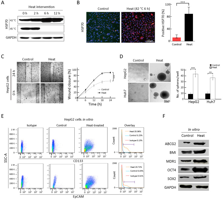
Figure 2.
Enrichment of TICs by heat intervention in vitro. (A,B) Western blot (A) and immunofluorescence analysis (B) of HSP70 expression by heat treatment of HepG2 cells. Nuclei were stained with DAPI; (C) Wound healing analysis for the closure ability of HepG2 cells after heat treatment; (D) Sphere-forming ability of HepG2 and Huh7 cells after heat treatment. The number of spheres more than 50 μm was quantified; (E) Proportions of CD133+ and EpCAM+ subpopulation of HepG2 cells in the indicated treatment groups were analyzed by FCA; (F) Western blot analysis for the stem cell-related genes expression of HepG2 cells in control and heat treatment group. Bar graphs in (B,D) are presented as
 . TIC, tumor-initiating cell; HSP, heat shock protein; DAPI, 4’,6-diamidino-2-phenylindole; EpCAM, epithelial cell adhesion molecule; FCA, flow cytometric analysis; SSC-A, side scatter area. **, P<0.01;***, P<0.001.

