FIG. 3.
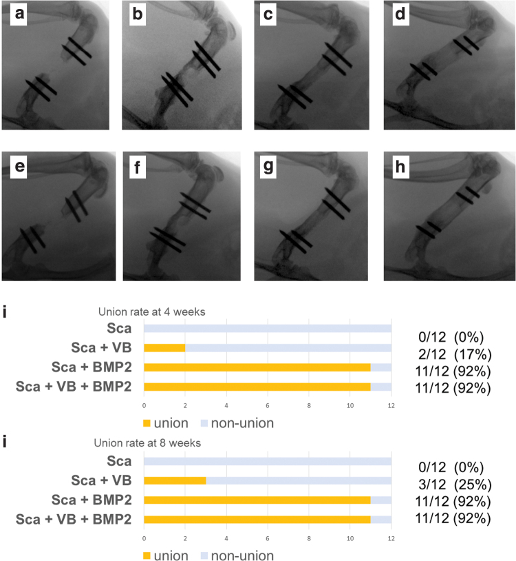
X-rays taken at 4 weeks (a–d) and 8 weeks (e–h) after surgery. (a) Almost no bone formation was seen in Sca Group at 4 weeks. (b) Bone formation along the scaffold was observed in Sca+VB Group at 4 weeks. Complete bone bridging over the defect was seen in Sca+BMP-2 Group (c) and Sca+VB+BMP-2 Group (d) at 4 weeks. (e) Bone volume in the defect area was small in Sca Group at 8 weeks. (f) Considerably more bone formation was seen in Sca+VB Group at 8 weeks. The greatest bone formation was observed in Sca+BMP-2 Group (g) and Sca+VB+BMP-2 Group (h). (i) Rate of bone union in the defect area at 4 weeks. (j) Rate of bone union in the defect area at 8 weeks. BMP-2, bone morphogenetic protein-2. Color images are available online.

