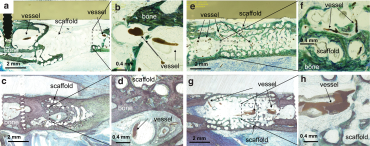
FIG. 7.
Histology images stained with Stevenel's blue and Van Gieson's picrofuchsin. (a, b) Sca Group (c, d) Sca+VB Group. (e, f) Sca+BMP-2 Group, (g, h) Sca+VB+BMP-2 Group. (b, d, f, h) High magnification images of area indicated by dashed boxes. (a, b) Only a small amount of bone was observed along the scaffold in Sca Group. The formation of vessel-like structures was limited around the interface of the femoral bone and scaffold. There was almost no vessel formation inside the scaffold. (c, d) Bone formation was seen around the scaffold in Sca+VB Group. A vessel-like structure filled with Microfil was seen inside the central tunnel. (e, f) Bone formation bridging the defect was seen in Sca+BMP-2 Group. Small vessel-like structures filled with Microfil were observed throughout the scaffold. (g, h) Bone was growing along the scaffold in Sca+VB+BMP-2 Group. A vessel-like structure was seen in the central tunnel in the scaffold. Color images are available online.

