Fig. 1.
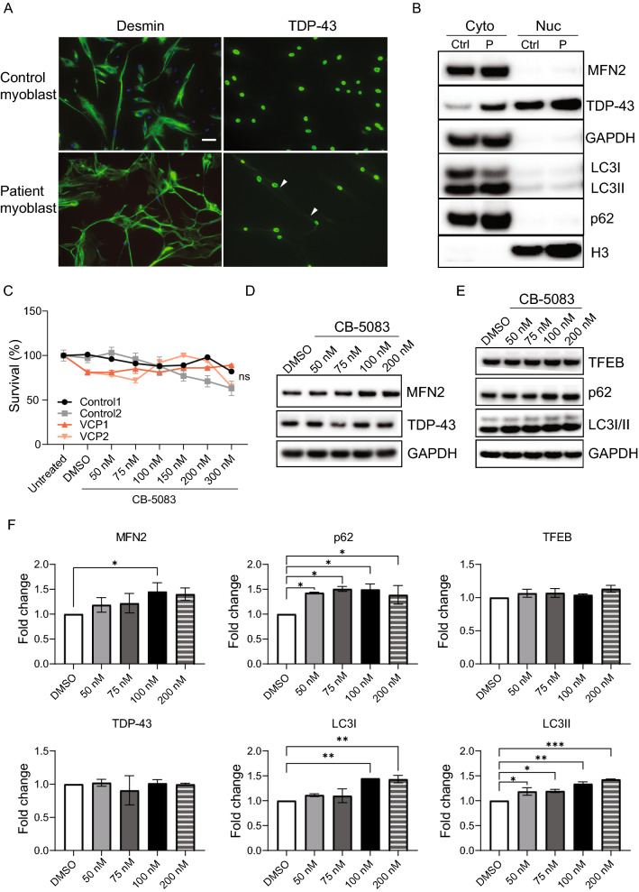
Evaluation of CB-5083 with patient-derived primary myoblasts. A Immunohistochemical analysis indicates that primary myoblasts express the myoblast marker desmin (green). TDP-43 cytoplasmic signaling (green) is present in patient myoblasts (scale = 20 µm, nuclear stain = blue). Arrowhead points to the cytoplasmic TDP-43 signals. B Analysis of nuclear-cytoplasmic fractions reveals that cytoplasmic p62 levels are higher in the patients’ myoblasts (P) compared to control myoblasts (Ctrl). GAPDH and H3 were used as loading controls for cytoplasmic and nuclear fractions, respectively. Western Blot experiments were repeated in two patients and two control myoblasts for two times, and the results were consistent. C Patient and control myoblasts tolerated serial treatment with CB-5083 at concentrations up to 300 nM. The MTT analysis was performed in two control and two patient myoblasts for three time. No significant differences were observed across the four samples (P = 0.557, one-way ANOVA, followed by Fisher’s LSD). D, E Dose–response effects of CB-5083 on biomarkers in patient myoblasts. Autophagy markers, LC3I/II and p62, and MFN2 were upregulated upon CB-5083 treatment. TDP-43 and TFEB did not change. F Quantification of Western blots from two independent patient myoblast lines was shown in (D, E). Western blot was repeated at least twice. Statistical analysis was performed by one-way ANOVA followed by Fisher’s LSD test. *P < 0.05. **P < 0.01. ***P < 0.001

