Fig. 1.
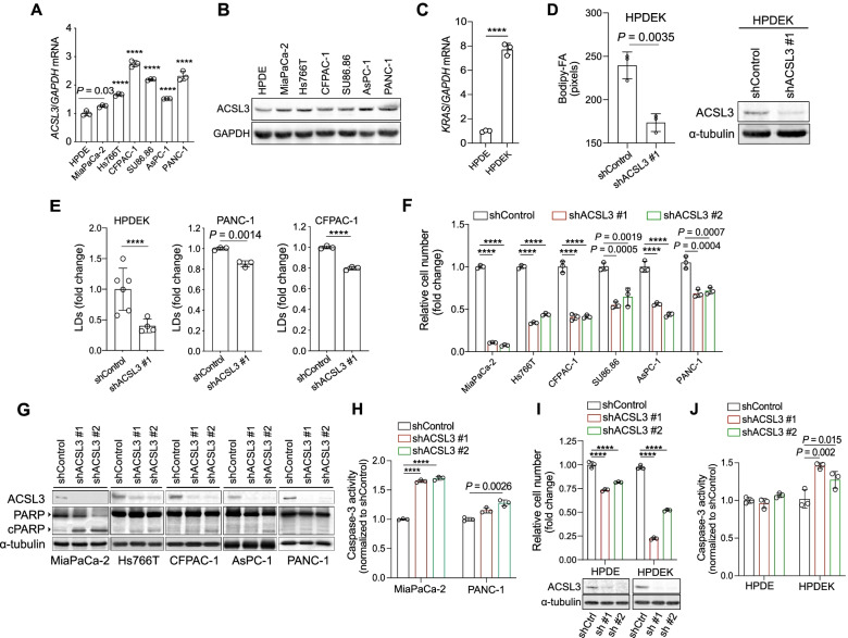
ACSL3 knockdown reduces the proliferation of PDAC cells. A and B ACSL3 mRNA (A) or protein (B) level in immortalized HPDE and the indicated human pancreatic cancer cell lines. HPDE: human pancreatic ductal epithelial cells; n = 3. C KRAS mRNA level of HPDE cells treated with 500 ng/ml doxycycline for 72 h to induce KRASG12D (HPDEK); n = 3. D Bodipy-FA uptake of HPDEK cells transduced either with an empty vector (pLKO-puro, shControl) or a shRNA against ACSL3 (shACSL3 #1), stained with BODIPY 500/510 C4, C9 and analysed by confocal fluorescence microscopy (left), and immunoblot showing ACSL3 knockdown efficiency (right). Statistical analysis was performed from ~ 150 cells/sample; n = 3. E Lipid droplets (LDs) staining with LipidTOX followed by flow cytometry quantification of HPDEK (n = 4–6), PANC-1 (n = 3) and CFPAC-1 (n = 3) cells transduced as in (D). F Relative cell number of the indicated cell lines transduced either with an empty vector (pLKO-puro, shControl) or 2 different shRNAs against ACSL3; n = 3. G Immunoblot analysis of the indicated cell lines transduced as in (F). H Caspase-3 activity of MiaPaca-2 and PANC-1 cells transduced as in (F) before plating for the assay; n = 3. I Relative cell number (top) and immunoblot to show ACSL3 knockdown efficiency (bottom) of HPDE and HPDEK cells transduced either with an empty vector (pLKO-puro, shControl) or 2 different shRNAs against ACSL3 and treated with 500 ng/ml doxycycline (HPDEK) for 72 h before plating for the assay; shCtrl: shControl, sh #1: shACSL3 #1, sh #2: shACSL3 #2; n = 3. J Caspase-3 activity of HPDE cells transduced and treated as in (I); n = 3. Graphical data are shown as the mean ± s.d. Statistical analyses were done using two-tailed unpaired Student’s t-test or one-way ANOVA. **** P < 0.0001; n, number of biologically independent samples

