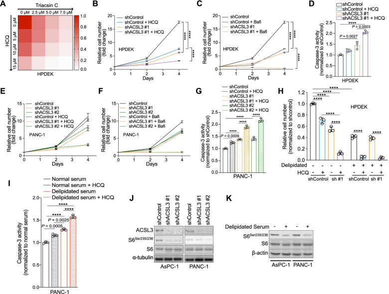
Fig. 5.
Concomitant extracellular lipid depletion and autophagy targeting strongly affect PDAC cell viability. A Heatmap obtained by measurement of HPDEK cell number 96 h upon treatment with incremental combined doses of Triacsin C and HCQ. HCQ: hydroxychloroquine. B and C Relative cell number of HPDEK cells transduced with a shRNA against ACSL3 (shACSL3 #1) and treated with 10 μM HCQ (B) or 100 nM bafilomycin (C). HCQ: hydroxychloroquine. Bafi: bafilomycin; n = 3. D Caspase-3 activity of HPDEK cells treated as in (B). HCQ: hydroxychloroquine; n = 3. E and F Relative cell number of PANC-1 cells transduced with 2 different shRNAs against ACSL3 and treated with 10 μM HCQ (E) or 100 nM bafilomycin (F). HCQ: hydroxychloroquine. Bafi: bafilomycin; n = 3. G Caspase-3 activity of PANC-1 cells treated as in (E). HCQ: hydroxychloroquine; n = 3. H Relative cell number of HPDEK cells transduced with a shRNA against ACSL3 (sh #1: shACSL3 #1) and treated as indicated for 72 h. HCQ: hydroxychloroquine; n = 3. I Caspase-3 activity of PANC-1 cells treated with media containing normal or delipidated serum for 72 h and/or 10 μM HCQ. HCQ: hydroxychloroquine; n = 3. J and K Immunoblot analysis for the indicated targets upon ACSL3 knockdown with 2 different shRNAs (J) and upon treatment with media containing delipidated serum for 72 h (K). Graphical data are shown as the mean ± s.d. Statistical analyses were done using one- or two-way ANOVA. **** P < 0.0001; n, number of biologically independent samples

