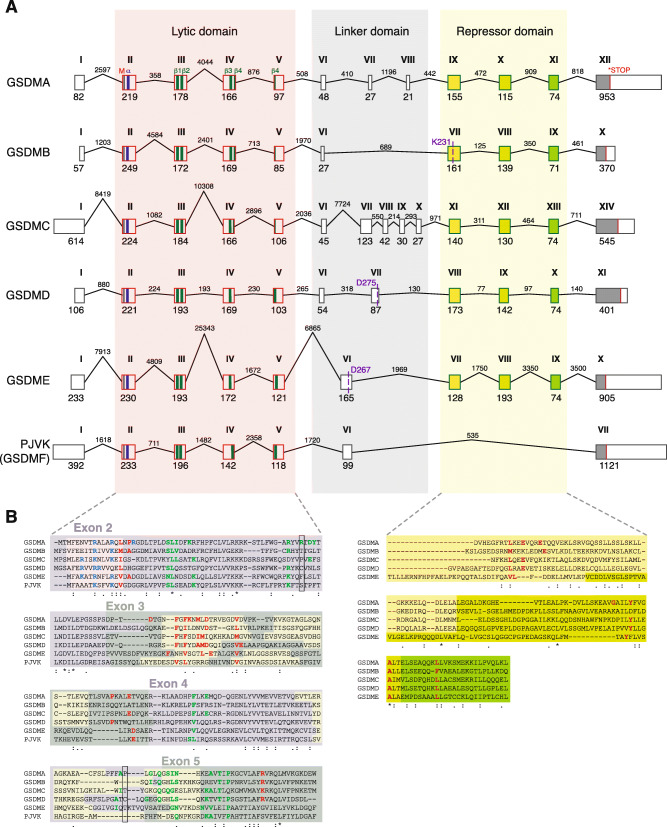
Fig. 5.
Analysis of the exons and introns present in human gasdermin genes. A Schematic representation of the different exons and introns present in the genomic DNA of the different human gasdermins. The first methionine is indicated with a black line, the stop codon with a red line, the first α-helix of the N-terminal domain is indicated with a blue bar, and the four β-sheets that integrate into membranes are indicated with green bars. The exons of the repressor C-terminal domain are represented with different colors to show the conserved similarity between them. Cleavage sites for granzyme A (K231) in human GSDMB, caspase-1 (D275) in human GSDMD and caspase-3 (D267) in human GSDME are shown. For human gsdmb, the gsdmb-403 splice variant is shown. B Protein alignment of the N-terminal and repressor C-terminal domains with the different exons highlighted. The residues that are part of the α-helix are highlighted in yellow, and the positive residues involved in the lipid binding are presented in blue. The residues involved in the insertion into membranes are presented in green and the residues involved in the oligomerization of different subunits to form the pore are presented in red. The positions of the Cys51 and Cys191 of human GSDMD are shown by a black box. In the C-terminal, the residues presented in red are important for the auto-inhibition

