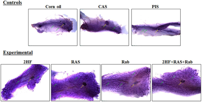
Figure 1. Whole mount preparations of mammary gland fat pads from SENCAR mice.
Mammary gland fat pads, excised from DMBA-administered SENCAR mice that received a control treatment (A, corn oil; B, CAS; C, PIS) or an experimental treatment (D, 2HF; E, RAS; F, Rab; G, a combination of 2HF+RAS+Rab), and stained with Mayer’s hematoxylin. Mammary gland fat pads from the control treatment groups were slightly small and edematous, making it difficult to achieve adequate spread on glass slide. The lymph nodes associated with the fat pads were present in all groups (yellow arrow). The development of the duct system (green arrows) was markedly attenuated in all control treatment groups when compared with any of the experimental treatment groups. Fat pads in all of the experimental treatment groups are similar to each other. The stained glands were placed in 70% ethanol until photographed using a Leica MZ 10F Stereomicroscope (Chicago, IL). Abbreviations: 2HF, 2'-hydroxyflavanone-treated; CAS, scrambled antisense-treated; RAS, RLIP antisense-treated; PIS, preimmune serum-treated; Rab, RLIP antibody-treated.

