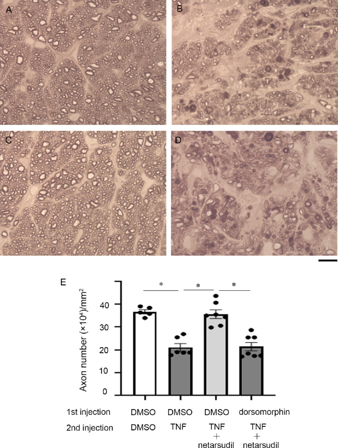
Figure 8.
Light microscopic study of PPD-stained axons 2 weeks after the second intravitreal administration of (A) DMSO, (B) TNF, or (C, D) 200 pmol netarsudil + TNF. First injection of A to C DMSO or D 200 pmol dorsomorphin was performed 1 hour before the second injection. Scale bar: 10 µm. (E) Quantification of axon number; n = 5 to 7 per experimental group. *P < 0.0001.

