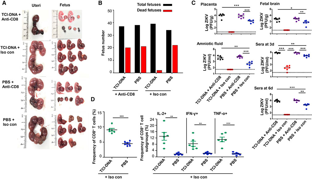
Figure 7. CD8+ T cell immune responses induced by ZIKV TCI-DNA vaccine played a key role in protecting pregnant mothers and their fetuses against ZIKV infection.
Female BALB/c mice were immunized with ZIKV TCI-DNA or PBS control for two doses and mated with male BALB/c mice for pregnancy at 10 days post-second immunization. The pregnant (E10–E12) mice were then injected (i.p.) with anti-CD8a (for depleting CD8+ T cells) or IgG2a isotype control (i.e., Iso con; without depleting CD8+ T cells) antibody (200 μg/mouse) for three times (−2, −1, and 3 days p.i.). One day before challenge, the mice were also injected with anti-IFNAR1 blocking antibody (for depleting type I IFN; 2 mg/mouse) and then infected with ZIKV (strain R103451, 106 PFU/mouse).
(A–C) Six days post-challenge, the mice were euthanized, recorded for morphology of uteri and fetuses (A), and counted for dead and total fetuses (B), and viral titers were determined by plaque-forming assay in placenta, amniotic fluid, and fetal brain, as well as sera collected at 3 and 6 days post-challenge (C). The detection limit was 20 PFU/g (for placenta), 40 PFU/g (for fetal brain), and 50 PFU/ml (for sera and amniotic fluid). Six days post-challenge, splenocytes were isolated from the mice injected with isotype control antibody (i.e., Iso con) and analyzed for ZIKV-specific CD8+ T cell responses by flow cytometry analysis.
(D) Quantification of the frequencies of CD8+ T cells (left), as well as IL2+, IFN-γ+, and tumor necrosis factor (TNF)-α secretion in CD8+ T cells (right) in splenocytes.
The data are represented as mean ± SEM (n = 6). *p < 0.05; **p < 0.01; ***p < 0.001.

