Figure 2.
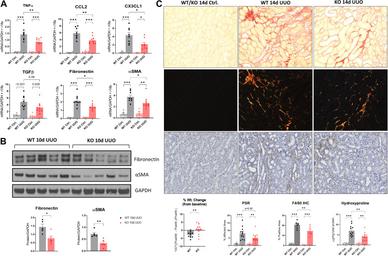
Genetic silencing of 15-lipoxygenase (15-LO) is protective during unilateral ureteral obstruction (UUO) progression. A: quantitative RT-PCR for tumor necrosis factor-α (TNF-α), monocyte chemoattractant protein-1 [chemokine (C-C motif) ligand 2 (CCL2)], fractalkine [chemokine (C-X3-C motif) ligand 1 (CX3CL1)], transforming growth factor-β (TGF-β), fibronectin, and α-smooth muscle actin (α-SMA) at 10 days (d) after UUO vs. respective controls (Ctrl) from wild-type (WT) and knockout (KO) animals. Data were normalized to the average of GAPDH + r18S. *P < 0.05, **P < 0.01, and ***P < 0.001 by one-way ANOVA. B: Western blots of fibronectin and α-SMA protein expression at 10 days after UUO. *P < 0.05 and **P < 0.01 by t test. C: fibrosis was quantified 14 days after UUO using picrosirius red (PSR) staining for collagen. Shown are representative ×20 light microscopic and corresponding polarized images (top), which were quantified according to the percent affected cortical area. The control specimens shown represent the typical appearance of WT or KO control kidneys. F4/80 immunohistochemistry (IHC) was performed on 14-day UUO specimens (bottom), and the corresponding quantification of the percent positive area is shown. *P < 0.05, **P < 0.01, and ***P < 0.001 by one-way ANOVA. Separately, hydroxyproline content for fibrosis was determined using a colorimetric assay from these same animals and then normalized to total protein concentration (bottom right graph). Animal weight changes at the time of euthanasia were also obtained (bottom left graph). **P < 0.01 by t test.

