Figure 1.
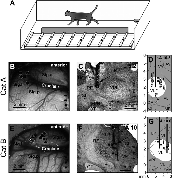
Locomotion tasks and reconstruction of recording sites in the motor cortex and injection sites in the ventrolateral thalamus (VL). A: the walking chamber was divided into two corridors. In one corridor, the floor was flat; the other corridor contained a horizontal ladder. Black circles on the crosspieces of the ladder schematically show placements of the right forelimb paw, whereas gray circles show those of the left forepaw. Sites of recording in the forelimb representation of the left motor cortex of cat A (B) and cat B (E). Microelectrode entry points into the cortex are shown as symbols of different shapes. □, penetrations where the majority of neurons had receptive fields (RFs) on the shoulder, arm, and/or forearm but not the wrist or paw; ◊, penetrations where most neurons had RFs on the arm and/or forearm and also responded to stimulation of the wrist and/or paw; Δ, penetrations where most neurons had RFs on the wrist and/or paw. Overlapping symbols designate penetrations in which different neurons had RFs on different segments of the forelimb. A circle in B shows a penetration in which only neurons without a somatosensory RF were recorded (two neurons). In E, the microelectrode track where neurons were recorded—the activity of which is shown in Figs. 3 and 6—is indicated by a white cross. C, D, F, G: sites of recording and injection in the VL. Parasagittal (C) and frontal (F) sections of the thalamus of cats A and B, respectively. Cresyl violet stain. Arrows point to electrolytic lesion marks. D and G: reconstruction of areas of 6-cyano-7-nitroquinoxaline-2,3-dione (CNQX) inactivation in cat A (D) and cat B (G). These are modified fragments from Fig. 2D from Ref. 29. Vertical black bars schematically represent injection cannulas. White circles highlight approximate areas affected by CNQX. Symbols indicate the positions of VL neurons whose activity was described by Marlinski et al. (29). Squares show neurons with somatosensory RFs on the shoulder; diamonds show cells that were activated by movements in the elbow; up-facing triangles indicate neurons with RFs on the wrist or paw; down-facing triangle shows a neuron whose RF encompassed the entire forelimb; and circles indicate neurons without somatosensory RFs and those where the RFs were not identified. Filled symbols represent neurons with axon projecting to the motor cortex; open symbols represent neurons whose axonal projections were not identified. AV, nucleus anteroventralis thalami; CI, capsula interna; CL, nucleus centralis lateralis; Cruciate, sulcus cruciatus; EPN, nucleus entopeduncularis; LA, nucleus lateralis anterior; LME, lamina medullaris externa thalami; LP, nucleus lateralis posterior; NC, nucleus caudatus; OT, optic tract; PC, pedunculus cerebri; RE, nucleus reticularis thalami; Sig.a., gyrus sigmoideus anterior; Sig.l., gyrus sigmoideus lateral; Sig.p., gyrus sigmoideus posterior; SUB, nucleus subthalamicus; VA, nucleus ventralis anterior; VL, nucleus ventralis lateralis; VM, nucleus medialis; VPL, nucleus ventralis posterolateralis.

