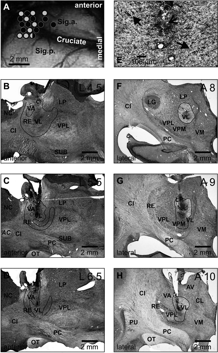
Figure 9.
Reconstructions of recording sites in the motor cortex and lesion sites in the ventrolateral thalamus (VL) of cat A. A: sites of recording in the forelimb representation of the left motor cortex. Microelectrode entry points into the cortex that were made before the lesions are shown by black circles, while those made after the lesions are shown by open circles. B–D: parasagittal sections of the left thalamus. Areas where most cells were destroyed by kainic acid (KA) are outlined. The circled areas in the VA appeared intact. E: high magnification photomicrograph of a section through the lesion site in the left thalamus. An arrow with an open head points to the KA injection site. Arrows with solid heads point to several surviving neurons next to the KA injection site. F–H: frontal sections of the right thalamus. Areas where most cells were destroyed by kainic acid are outlined. B–H: cresyl violet stain. AC, anterior commissure; AV, nucleus anterio-ventralis thalami; CI, capsula interna; CL, nucleus centralis lateralis; Cruciate, sulcus cruciatus; LG, lateral geniculate nucleus; LP, nucleus lateralis posterior; NC, nucleus caudatus; OT, optic tract; PC, pedunculus cerebri; PU, putamen; RE, nucleus reticularis thalami; Sig.a., gyrus sigmoideus anterior; Sig.l., gyrus sigmoideus lateral; Sig.p., gyrus sigmoideus posterior; SUB, nucleus subthalamicus; VA, nucleus ventralis anterior; VL, nucleus ventralis lateralis; VM, nucleus medialis; VPL, nucleus ventralis posterolateralis; VPM, nucleus ventralis posteromedialis.

