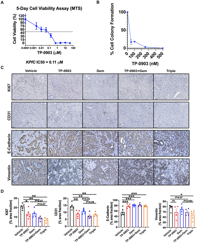
Figure 3. TP-0903 induces a differentiated tumor cell phenotype in KPfC GEMM model.
A) IC50 for TP-0903 in KPfC cells (BMF-A3) was determined by MTS assay. Response was validated in replicate plates (n ≥ 4). IC50 for BMF-A3 was determined to be 110 nM. B) BMF-A3 were plated at low density for colony forming assay. Cells were treated with DMSO or TP-0903 at 100, 250, 500 nM or 5 μM. After 10 days, cells were fixed with 10% formalin and stained with crystal violet. N> 3 for each dose. Colonies were counted and each dose was normalized to the control. C) Tumor tissues from KPfC mice treated with vehicle, TP-0903, gemcitabine, TP-0903+gemcitabine, TP-0903+gemcitabine+anti-PD1 were evaluated by IHC for Ki67, CD31, E-Cadherin and vimentin. Scale bar, 100 μm. Images were analyzed using ImageJ software. Quantification of % area fraction is shown in D). *, P<0.05; **, P < 0.01; ***P < 0.005; by ANOVA with Tukey’s MCT.

