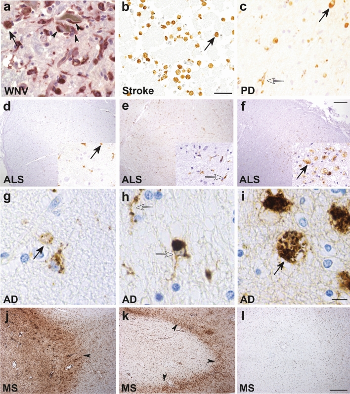
Fig. 1.
Macrophage/microglial CD68 tissue staining in human CNS pathologies. Ameboid (solid arrows, likely macrophages and/or reactive microglia) and ramified (open arrows, likely microglia) CD68+ myeloid cells are shown in various neuropathologies: a West Nile virus (WNV) [247]. Macrophage/microglia engulfing a degenerating neuron (arrowheads) in the substantia nigra in a patient with fulminant WNV encephalitis (400X magnification). b Cortical stroke [94]. Foamy macrophages/microglia are present in a cerebral infarct (several weeks old) (scale bar represents 50 μm). c Parkinson disease (PD) [65]. Ramified microglia and macrophages with enlarged cytoplasm and short stout processes are present in the substantia nigra (400X magnification). d–f Amyotrophic lateral sclerosis (ALS) [38]. The three images show the variable extent of microglia activation in the corticospinal tract in patients with ALS assessed as either mild (d), moderate (e) or severe (f) (scale bar represents 250 μm). g–i Alzheimer disease (AD) [148]. The three images show the rounded ameboid microglia (g), ramified microglia (h) or foamy macrophages (i) that can be seen in AD brains (scale bar represents 10 μm). j–l Multiple sclerosis (MS) [193]. Three images show variable inflammatory activity in MS, with either numerous foamy macrophages within a demyelinating plaque (j), macrophages at the rim of a plaque (arrowheads) (k), and an inactive plaque with only a few ramified microglia (l). All images reproduced with permission

