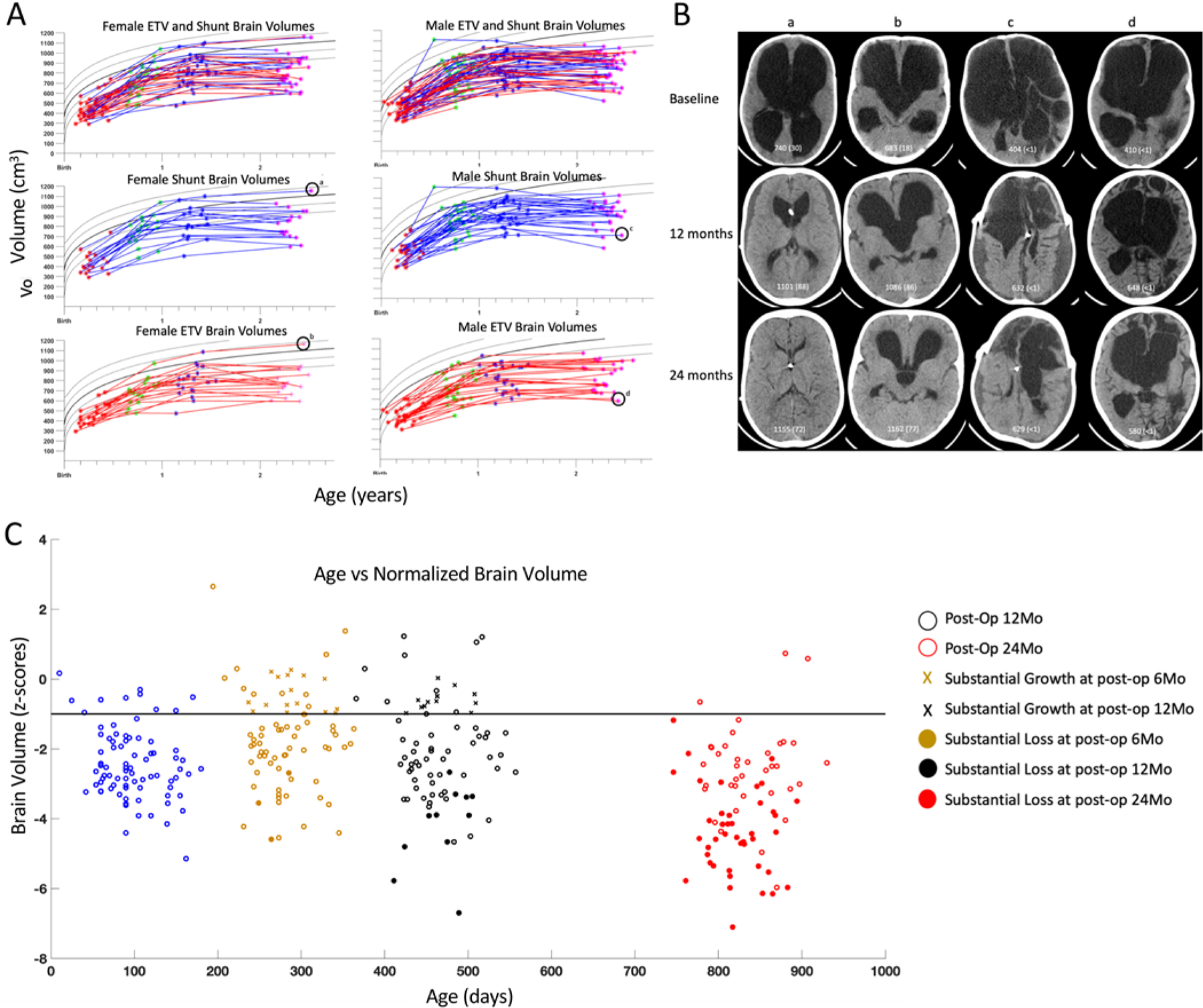
FIG. 3.

A: Raw brain volumes, in cm3, stratified by males, females, shunts, and ETV intention to treat. B: Representative CT images for cases of normal brain volume at 24 months for female shunt (a) and female ETV (b) and growth failure at 24 months for male shunt (c) and male ETV (d). Brain volume (and normalized percentage) is given on each image shown. C: Normalized brain volumes, age adjusted and plotted as z-scores, for volumes measured before surgery and 6, 12, and 24 months following surgery. Cases with substantial growth are indicated with x’s, and filled colored circles indicate substantial volume loss. The horizontal line at z = −1 indicates the threshold of normal age-adjusted volume (above) and volume less than normal for age (below).
