
Figure 1.
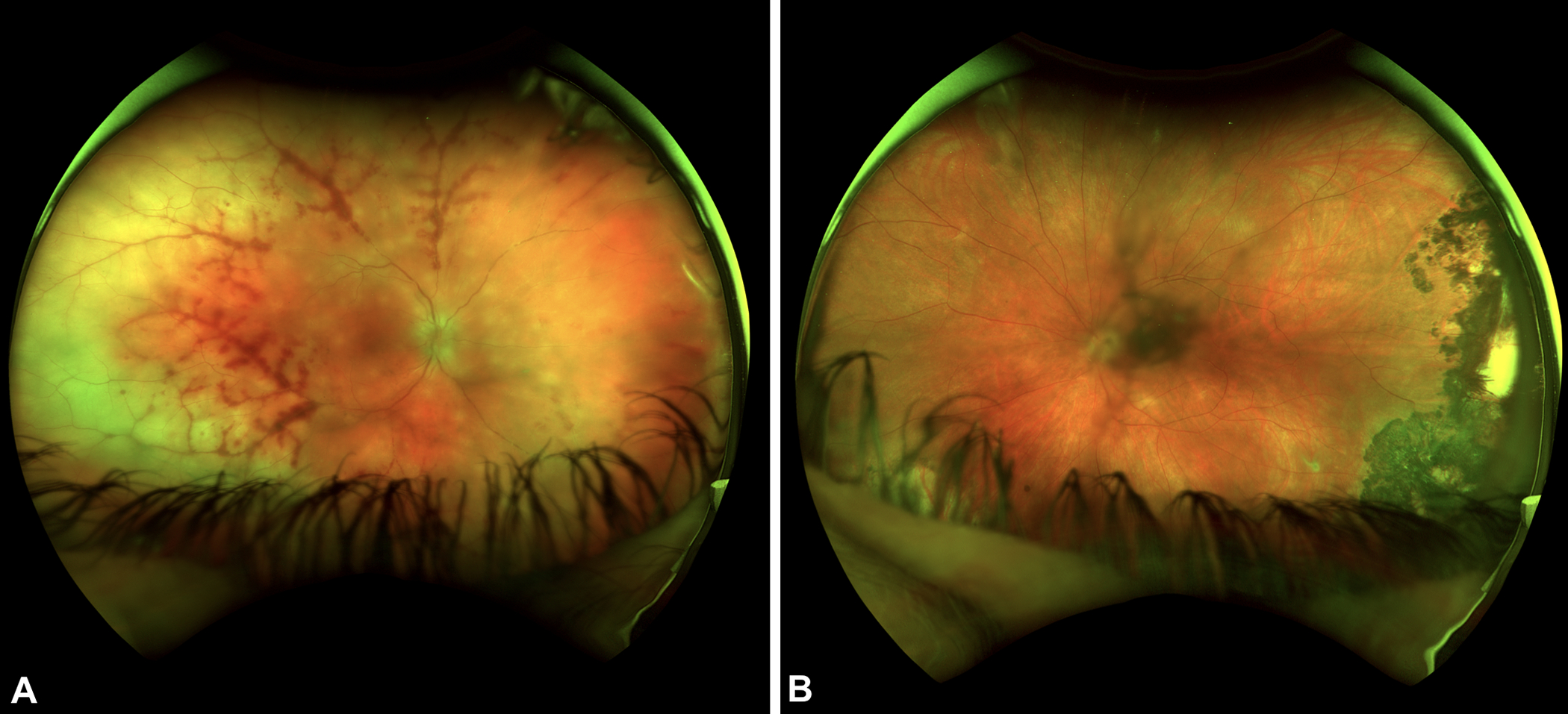
A) Ultra-widefield fundus photographs of patient 9 show diffuse peripheral retinal whitening temporally greater than nasally associated with severe perivascular hemorrhage. There are also multifocal areas of whitening in the peripapillary region and optic disc edema of the right eye. B) Ultrawide-field fundus photograph of the left eye shows a dense hyperpigmented chorioretinal scar temporally. She had a clinical diagnosis of toxoplasmosis previously but the extent of scarring suggests she may have had HSV ARN in the left eye previously.