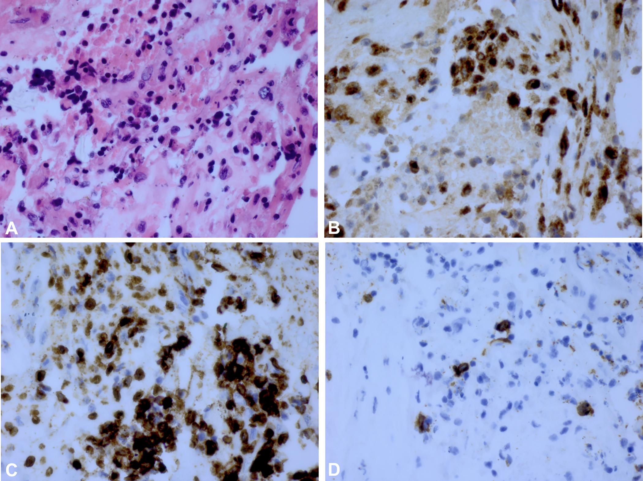
Figure 2.

A) H&E stain of vitreous biopsy for Case 1 (Patient 9) with HSV-2 associated ARN. B) Staining for CD68+ cells show a large number of macrophages in brown. C) The brown colored areas show a predominance of CD3+ T-cells. D) Staining for CD20+ B-cells shows scattered B-cells in brown suggesting a B-cell population within a predominantly T-cell lymphocyte population.
