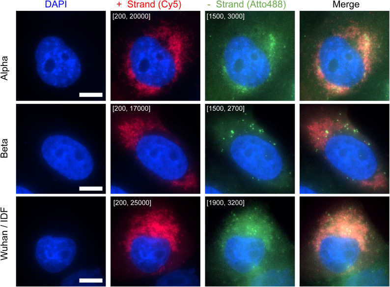
Figure S6. Imaging SARS-CoV-2 variants with CoronaFISH.
CoronaFISH images (with staining for the positive RNA) of Vero cells infected with SARS-CoV-2 variants alpha and beta and the original Wuhan/IDF strain. Cells were infected with an MOI of 0.1 and imaged 29 h post infection. Intensity scalings are indicated as described for Fig 1D. Scale bars: 10 μm.

