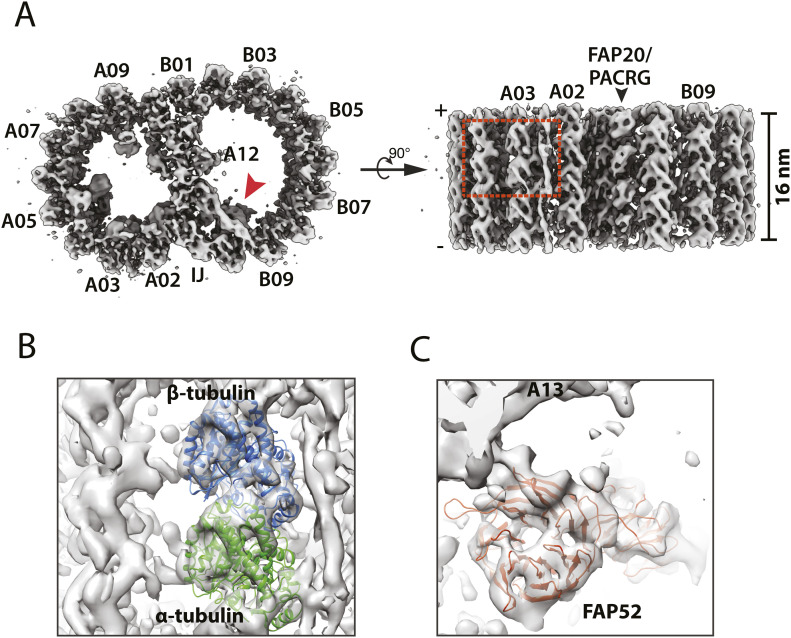
Figure 1. Structure of the 16-nm repeat of the doublet microtubule from the wild type.
(A) The DMT structure is displayed as iso-surface representation in two orthogonal views. Left: the DMT is viewed from the plus end. IJ: inner junction. Right: the DMT is viewed from the lumen of axoneme. (B) A magnified view of the boxed region in (A) shows that the secondary structure elements can be resolved in many parts of the map. Atomic models of α (green) and β (light blue) tubulin are fit into the protofilament A03 in the map. (C) An atomic model of FAP52, one of the microtubule inner proteins at the inner junction whose location in the DMT is indicated by a red arrowhead in (A), is fit into the average map. The blades of the β-propeller fold from the FAP52 WD40 domain are resolved in the map.

