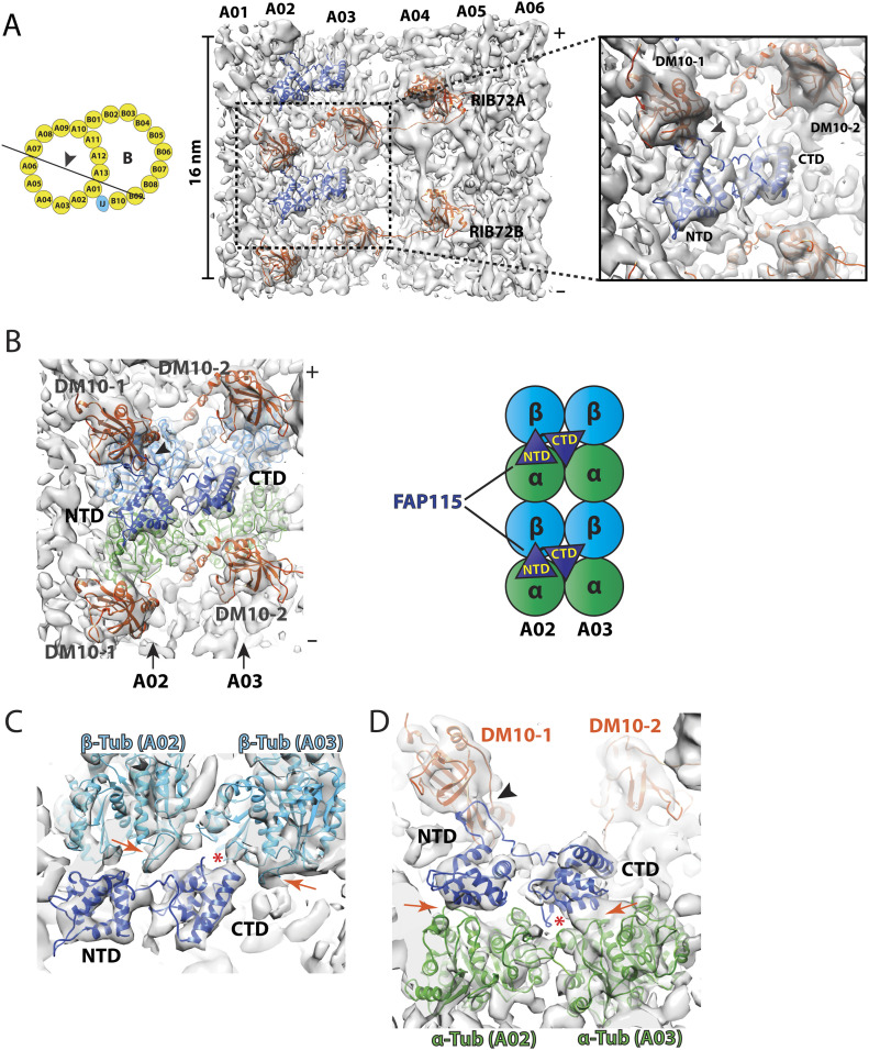
Figure 3. Binding of FAP115 to the luminal wall of doublet microtubule.
(A) FAP115 binds to protofilament A02 and A03 in the lumen of A-tubule. The atomic models for FAP115 (dark blue) and RIB72A/B (red) are fit into the density map. The viewing direction of the map is depicted in a cartoon on the left. The inset on the right shows a magnified view of FAP115. An arrowhead in the inset indicates the potential interaction between an extended loop from FAP115 and the first DM10 domain (DM10-1) from RIB72A that is anchored at protofilament A01. (B) Binding of FAP115 to protofilament A02 and A03. The density map is shown in the same viewing direction as in (A). Two pairs of α/β tubulin dimers are fit into the density map, α-tubulins are in green and β-tubulins are in light blue. The two EF-hand domains from FAP115, the N-terminal domain (NTD) and the C-terminal domain (CTD), are in dark blue. The cartoon illustrates the binding of FAP115 to two pairs of α/β tubulin dimers from pf A02/A03. (C) Potential interactions between FAP115 and two β-tubulins from pf A02/A03. The H1S2 loops from two β-tubulins are indicated by the red arrows. These two loops resemble two jaws of a vernier caliper where FAP115 CTD fits in and makes contacts. A red asterisk indicates potential interaction between FAP115 CTD and the S9-S10 loop from A03 β-tubulin, Cα-Cα distance < 7 Å. (D) Potential interactions between FAP115 and two α-tubulins from pf A02/A03. The two red arrows indicate potential interaction interface between FAP115 NTD, CTD and two α-tubulins, Cα–Cα distance < 7 Å. A red asterisk indicates an EF-hand loop from FAP115 that extends towards the lateral interface between two α-tubulins, making potential interactions.

