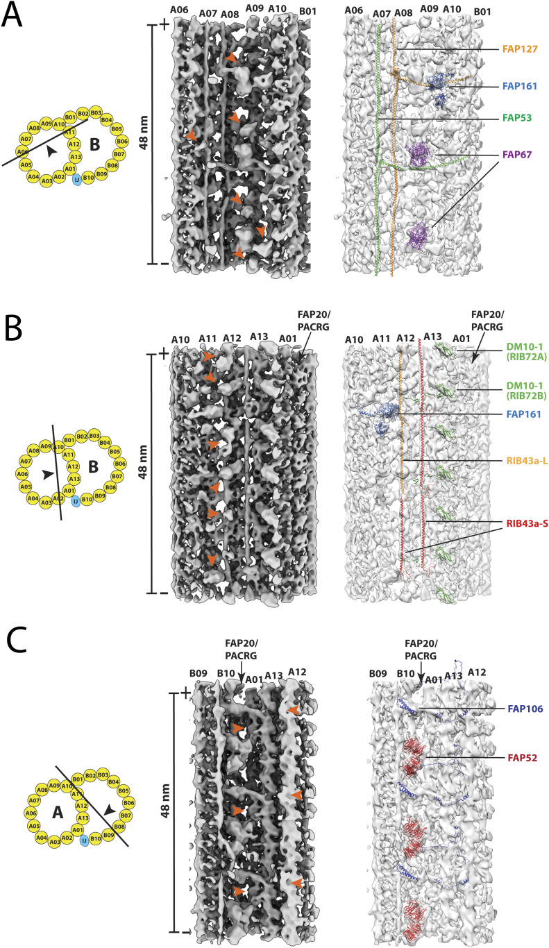
Figure 5. Structure comparison of the doublet microtubule between Tetrahymena and Chlamydomonas.
Three regions in the DMT are compared. These are as follows: (A) the A-tubule “seam” region spanning the pf A06∼A10, (B) the ribbon region, and (C) the inner junction region. In each panel, left: a cartoon depicts the viewing direction of the structures in the DMT; middle: the red arrowheads indicate the unidentified microtubule inner protein densities that are unique to the Tetrahymena DMT; right: the microtubule inner proteins found in both organisms are indicated. The atomic models from Chlamydomonas are fit into the Tetrahymena density maps in light gray.

