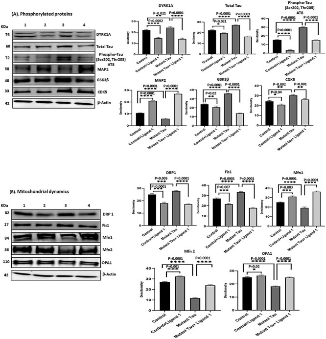
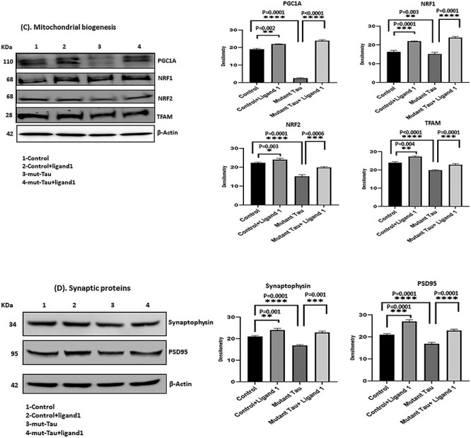
Figure 4.


(A) Immunoblotting analysis of phosphorylated tau ligand inhibition on DYRK1A, total Tau, PHF-Tau (AT8), MAP2, GSK3β and CDK5. Immunoblotting analysis was conducted using protein lysates from four group of mouse hippocampal cells (HT22). 1) HT22 cells (Control); 2) Control +ligand 1; 3) mutant tau (Tau cDNA transfected HT22) and 4) mutant Tau+ligand 1. 1) comparison with control versus control +ligand 1 treated cells significantly decreased levels of DYRK1A, total tau, PHF-Tau (AT8), GSK3β and CDK5 proteins were found with (P = 0.0002, P = 0.0021; P = 0.0001, P = 0.0001, P = 0.02, P = 0.04) and increased MAP2 (P = 0.0001); 2) comparison with mutant tau versus mutant tau + ligand 1 treated cells, significantly reduced levels of DYRK1A, total tau and PHF-tau (AT8), and GSK3β proteins were found with (P = 0.0001, P = 0.0001; P = 0.0001, P = 0.0001, P = 0.001) and increased MAP2 (P = 0.0001). (B) Immunoblotting analysis mitochondrial dynamics proteins. Increased levels of fission proteins Drp1 and Fis1 were observed in mutant tau (Tau cDNA transfected HT22; P = 0.005, P = 0.0001) when compared with Control. Reduced Drp1 and Fis1 levels were found (ligand 1 treated control cells (P = 0.0001, P = 0.007) and mutant tau+ligand 1 treated cells (P = 0.0001; P = 0.0001). Whereas mitochondrial fusion proteins Mfn1, Mfn2 and Opa1 were decreased in mutant Tau (P = 0.0001, P = 0.0001, P = 0.0001), when compared with control, increased in mutant tau+ligand 1 (P = 0.0001, P = 0.0001, P = 0.0001). (C) Immunoblotting analysis of mitochondrial biogenesis proteins. We found decreased levels of biogenesis proteins PGC1a, NRF1, NRF2 and TFAM in cells mutant tau when compared with control (P = 0.0001, P = 0.001; P = 0.0001, P = 0.0001, P = 0.0001). Increased PGC1a, NRF1, NRF2 and TFAM levels were found in cells treated with ligand 1 (control+ ligand 1) when compared with control (P = 0.002, P = 0.0001, P = 0.003, P = 0.004) and ligand 1 treated mutant tau cells (P = 0.0001, P = 0.0001, P = 0.0006, P = 0.0001). (D) Immunoblotting analysis of synaptic proteins Synaptophysin and PSD95. We found decreased levels of synaptic proteins synaptophysin and PSD95 in cells transfected with mutant tau cells when compared with control (P = 0.0001, P = 0.0001). Synaptic proteins were increased in mutant Tau+ligand 1 cells P = 0.001; PSD95, P = 0.0001) when compared with mutant tau cells.
