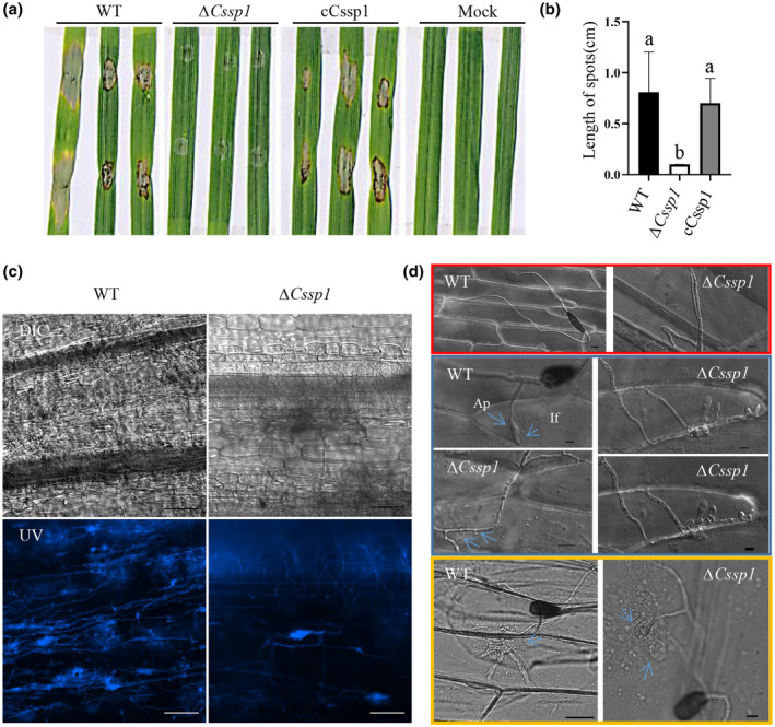
FIGURE 4.

Pathogenicity tests. (a) Detached leaves from Aikang 58 seedlings in a tray were inoculated with drops of a spore suspension (3 × 10⁴/ml) of wild type (WT), deletion mutant ΔCssp1, complemented strain cCssp1, or double deionized water as a mock control. The tray was kept in a moist chamber under darkness for 1 day and then moved to a greenhouse under a 16 h light/8 h dark photoperiod for 2 days. (b) Lesion lengths on wheat leaves were measured at 3 days postinoculation. Different letters indicate significant differences calculated by Tukey's LSD, p < 0.05. The experiments were repeated three times. (c) Mycelial expansion in barley leaves at 24 h after inoculation with fungal agar blocks. Fluorescent 7‐GFE staining was performed as described in Experimental Procedures. Bars, 100 μm. (d) Inner epidermis of onion bulbs 24 h after being inoculated with drops of spore suspensions (5 × 10⁴ cells/ml). Ap, appresorium; If, invasive hypha. Bars, 20 μm
