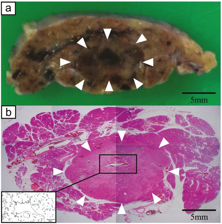
Figure 3.
a, Macroscopic findings of a resected specimen revealed that the tumor in the body of the pancreas formed a round and well-defined mass (white arrowheads). b, Hematoxylin-eosin staining showed that small lobules of the pancreas aggregated and morphologically exhibited nodular formation similar to a hyperplastic pancreatic lobule (white arrowheads). Inset: CK7 staining revealed that the central cyst-like lesion was an expanding pancreatic duct.

