
Fig. 3. False-positive diagnosis using the conventional criterion for anastomosis site abnormality in a 46-year-old male who underwent deceased-donor liver transplantation.
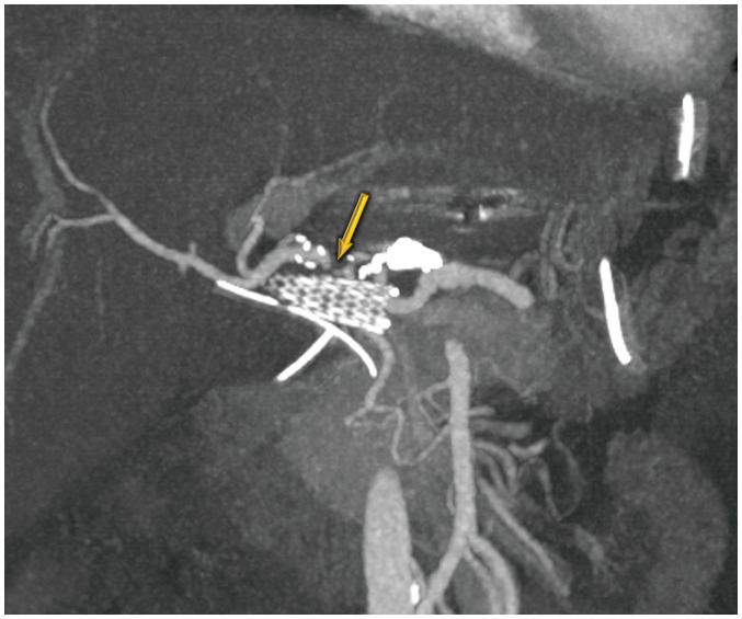
Maximal intensity projection image shows more than 50% focal narrowing (arrow) without distal run-off abnormality because of hepatic artery angulation. Doppler ultrasound abnormalities were normalized after 1 month, and no associated complication was seen in this patient within 6 months of follow-up.