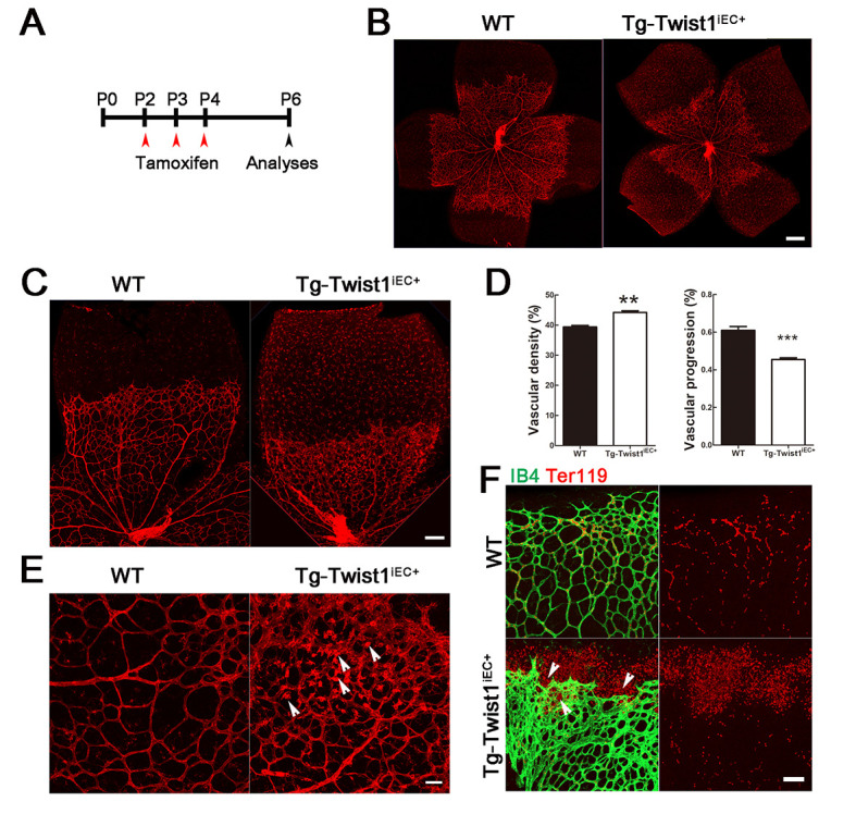
Figure 3.

Defective retinal angiogenesis and pathological NV in Tg-Twist1iEC+ mice at P6
A: Tg-Twist1iEC+ and control mice were intraperitoneally injected with tamoxifen at P2 to P4 and analyzed at P6. B–D: Isolectin B4 staining of retinal whole mounts showed delayed superficial vascular progression and increased vascular density in retinas of Tg-Twist1iEC+ mice compared with littermate WT mice. Scale bar: 250 μm for B; 100 μm for C; E: Arrows indicate aneurysmal-like pathological NV in Tg-Twist1iEC+ retinas. Scale bar: 50 μm. F: Red blood cells stained with Ter119 antibody show vessel leakage in Tg-Twist1iEC+ retinas, indicated by arrows. **: P<0.01; ***: P<0.001; two-tailed t-test. Scale bars: 50 μm.
