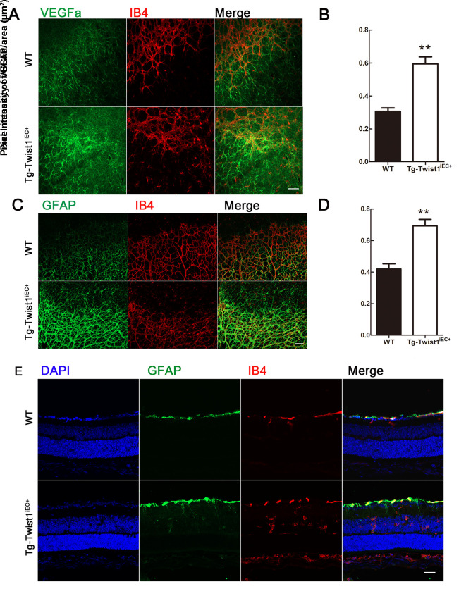
Figure 5.
Increased VEGFA secretion and enhanced gliosis inTg-Twist1iEC+ mice at P8
A–D: VEGFA (A, B), GFAP (C, D) and IB4 staining at vascular front in WT and Tg-Twist1iEC+ mice. VEGFA, green; GFAP, green; IB4, red. Scale bars: 50 μm. **: P<0.01, two-tailed t-test. E: Retinal section staining of GFAP expression to deep retina and INL. RGC: Retinal ganglion cell; INL: Inner nuclear layer; ONL, Outer nuclear layer. Scale bar: 25 μm.

