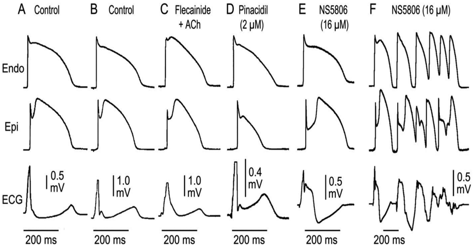
Figure 4.

Electrocardiographic manifestations of early repolarization recapitulated in coronary perfused canine ventricular wedge preparations. Each panel shows action potential recordings from epicardium (Epi) and endocardium (Endo) and a transmural ECG. J waves are a reflection of early repolarization of ventricular epicardium and can manifest as a J point elevation (A), a distinct J wave (B), a slurring of the terminal part of the QRS recorded following exposure to flecainide + acetylcholine (C ), a distinct J wave with an ST segment recorded following exposure to pinacidil (D), a gigantic J wave appearing as an ST segment elevation recorded following exposure to the Ito agonist NS5806 (E). It is under these conditions that we observe the development of polymorphic VT (F). Modified from (134) with permission.
