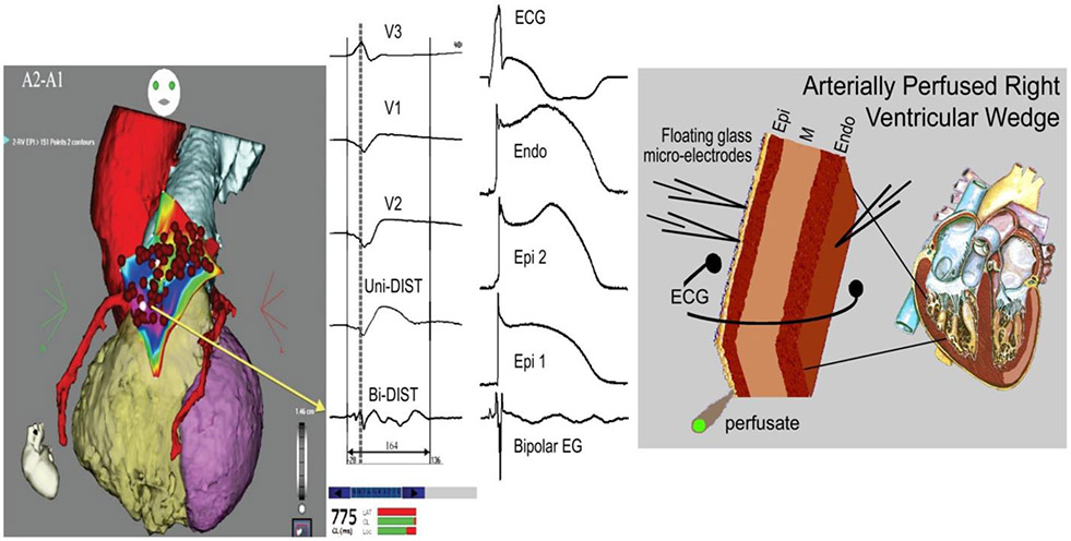
Figure 7.
Cellular basis for the appearance of fractionated bipolar electrograms (EG). Accentuation of the action potential notch and delay in the appearance of the action potential plateau (2 nd upstroke) gives rise to fractionated epicardial electrogram in the setting of Brugada syndrome (BrS). Left panel: Shown are right precordial lead recordings, unipolar and bipolar EGs recorded form the right ventricular outflow tract of a BrS patient (from Nademanee et al. (11), with permission). Right panel: ECG, action potentials from endocardium (Endo) and two epicardial (Epi) sites, and a bipolar epicardial EG (Bipolar EG) all simultaneously recorded from a coronary-perfused right ventricular wedge preparation treated with NS5806 (5 μM) and verapamil (2 μM) to induce the Brugada phenotype. Basic cycle length=1000 ms. Reproduced from reference,(81) with permission.

