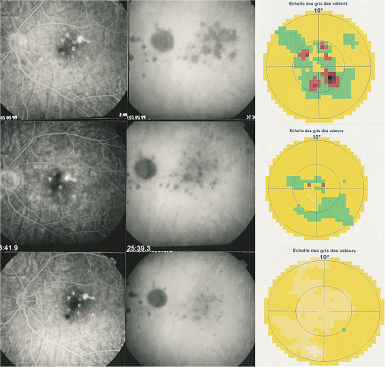
Fig. 12.
Correlation between FA, ICGA and visual field during corticosteroid treatment of MFC. This myopic patient presents with left eye photopsias, left decrease of visual acuity (VA) and the presence of a subjective left scotoma. FA (top left) only shows cicatricial FA lesions corresponding to cicatricial fundus lesions seen on fundus examination. ICGA (top middle) shows widespread hypofluorescent areas indicating fresh choriocapillaris lesions with corresponding visual field defects (top right). During corticosteroid therapy, practically no change is seen on FA, whereas ICGA signs of active disease progressively resolve together with visual field improvement (right column). (Reprinted from Diagnostics (Basel). 2021 May 24;11 (6):939. 10.3390/diagnostics11060939)

