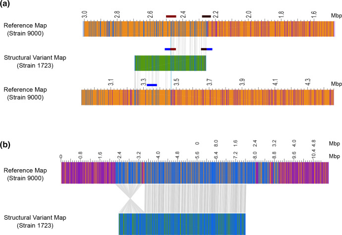
Fig. 5.
Optical mapping of E. coli O157:H7 PT21/28 strain 9000 variant Z1723. Structural variant (SV) analysis identified a 220 Kb duplication (a) and 1.2 Mb inversion (b) in the population of Z1723 relative to the reference strain 9000. The genome map (orange) of reference strain 9000 and each Z1723 structural variant (green) are shown. All restriction sites used to generate maps are shown. Paired restriction sites (blue lines) are aligned between the reference and variant maps (grey lines). Unpaired restriction sites (purple lines) outside aligned regions are also shown. The SV map containing the 220 Kb duplication has been aligned to two reference strain 9000 genome maps to demonstrate the hybrid composition of the map containing ΦStx2c at 3.4 Mb and an inverted 220 Kb duplicated region originating from between 2.2 and 2.4 Mb. The position of phage regions flanking the 220 kbp duplication (maroon and black blocks) and ΦStx2c (blue block) into which the duplication has inserted are indicated.

