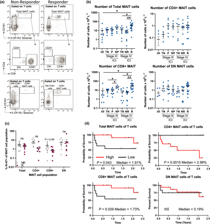
Figure 4.

The frequency of total and CD8+ MAIT cells is positively associated with improved clinical responses and overall survival in melanoma patients. (a) Example staining of circulating MAIT cell subsets in responding vs. non‐responding melanoma patients. (b) Comparisons of the absolute number of total, CD4+, CD8+ and double‐negative (DN) MAIT cell populations in early stage (I/II, n = 16), stage III (n = 25) [treatment naïve (TN), progressed on treatment (P), did not progress on therapy (NP), immune checkpoint inhibitor (ICI)] or stage IV (n = 37) [treatment naïve (TN), non‐responding (NR), responding (R), immune checkpoint inhibitor (ICI)]. (c) Comparisons of the frequency of proliferating cells (Ki‐67+) in anti‐PD‐1 non‐responders (NR, n = 10) compared to responders (R, n = 10). (d) Kaplan–Meier curves comparing the survival of stage IV melanoma patients who received at least three doses of anti‐PD‐1 therapy (n = 27) based on the median frequency of the listed MAIT cell population. The median for each cellular population is indicated on each graph. Clinical characteristics for all patients in this analysis can be found in Supplementary tables 2–5. All treated patients received at least three doses of ICI prior to sample collection. Survival was calculated from the time of the blood draw and all blood draws occurred, while the patients were still receiving therapy. Response to ICIs was characterised using RECIST1.1 criteria with responders identified as complete response (CR) or partial response (PR), while non‐responders were identified as stable disease (SD) or progressive disease (PD).
