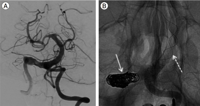
Fig. 3.

(A) Pre-treatment DSA image demonstrates the diseased right vertebrobasilar segment. (B) Image after deployment of the LEO stent showing the coiled right V4 segment after the 1st treatment (solid white arrow) and the LEO stent placement (dashed white arrow) during the 2nd treatment for a progressively dilating diseased lower basilar segment. DSA, digital subtraction angiogram.
