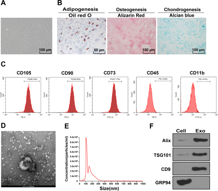
Fig. 2. Isolation and identification of ADMSCs and ADMSC-exosomes.
A The morphological characteristics of primary ADMSCs were observed under the inverted microscope. B Adipogenic (left), osteogenic (middle), and chondrogenic (right) differentiation experiments of ADMSCs. C The surface markers of ADMSCs were detected using flow cytometry. D The diameter of ADMSC-exosomes was observed under the TEM. E The diameter distribution and concentration of exosomes were detected using NTA. F The exosome marker proteins (Alix, CD9, and TSG101) and endoplasmic reticulum protein (GRP94) were detected using Western blot. The cell experiment was repeated three times independently.

