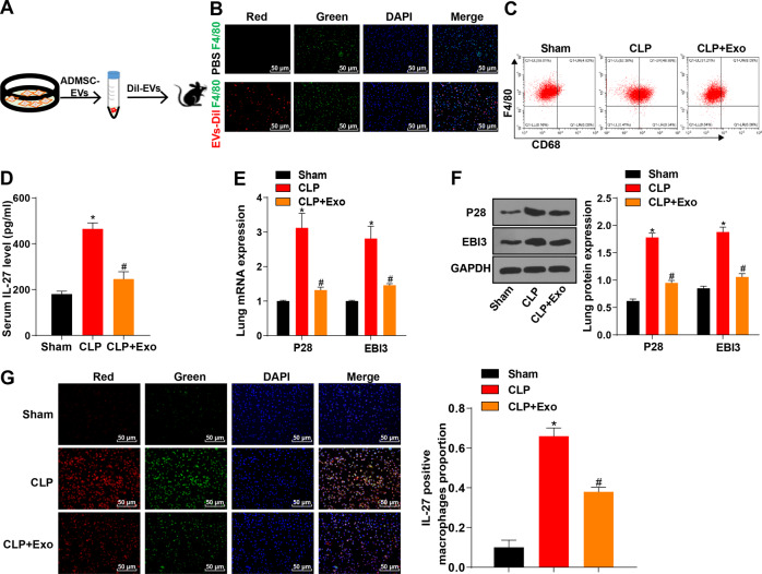
Fig. 4. ADMSC-exosomes reduced the number of pulmonary macrophages and the release of IL-27 in sepsis mice.
A ADMSC-exosomes were isolated, purified and labeled with Dil dye; flow chart of injection in CLP sepsis mice. B The co-localization of exosomes (Dil pre-labeled red) and macrophages (F4/80, green) in lung tissue was detected using immunofluorescence staining, scale bar = 25 μm. C The number of macrophages CD68+ F4/80 + in single-cell suspension of whole lung tissue was detected using flow cytometry. D The content of IL-27 in serum of CLP mice treated with ADMSC-exosomes was detected using ELISA. E The mRNA expressions of P28 and EB13 in lung tissue of CLP mice treated with ADMSC-exosomes were detected using RT-qPCR. F The protein levels of P28 and EB13 in lung tissue of CLP mice treated with ADMSC-exosomes were detected using Western blot. G The content of IL-27 in macrophages after the treatment of ADMSC-exosomes or not detected using immunofluorescence. Blue fluorescence indicated DAPI, green fluorescence indicated F4/80, and red fluorescence indicated IL-27. N = 8. Measurement data are depicted as mean ± SD. One-way ANOVA was employed for the comparisons among multiple groups, followed by Tukey’s multiple comparisons test. *p < 0.05 vs. Sham group; #p < 0.05 vs. CLP group.

