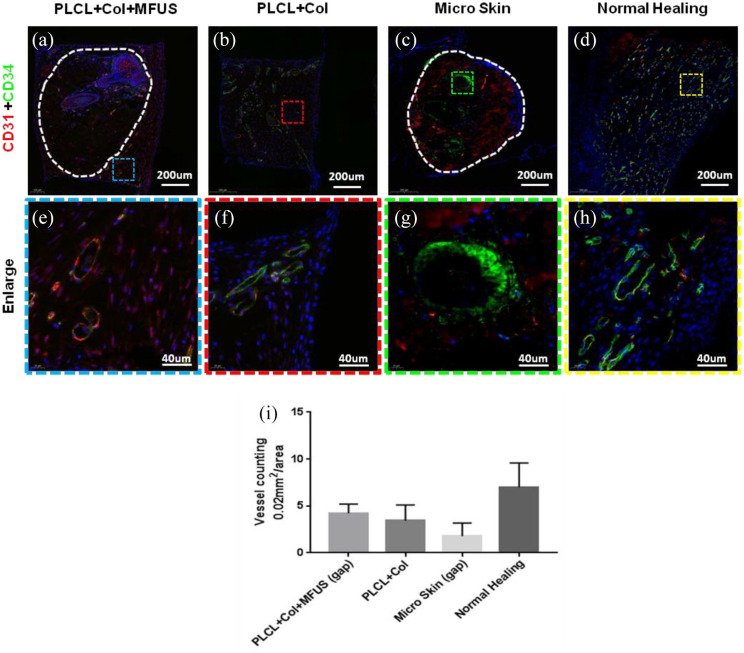
Figure 7.
Expression of blood vessel markers, CD31 and CD34 on rat dorsal skin tissue sections with four different treatments at day-7 by immunostaining. (a and e) The wound treated with tissue engineering skin (PLCL + Col + MFUS) showed that the vessels not only grew in the MFUS, but also grew in the gap areas (blue box areas) between the MFUSs and the PLCL scaffold. (b and f) The wound treated with PLCL + Col showed many positively stained blood vessels with CD34. (c and g) The wound treated with micro skin islands showed that a few blood vessels in the outside areas of MFUS were positively stained with CD31 and CD34, but some blood vessels in the inside of MFUS were positively stained with CD31 and CD34. (d and h) The wound without treatment (Normal healing) showed that many blood vessels were positively stained with CD31 and CD34. (i) Semi-quantification of the immunostaining results showed that the density of the blood vessels was 4.22/0.002 mm2 in PLCL + Col + MFUS treated wound, 3.44/0.002 mm2 in PLCL + Col treated wound, 1.78/0.002 mm2 in micro skin treated wound, and 7.01/0.002 mm2 in normal healing wound.

