Figure 8.
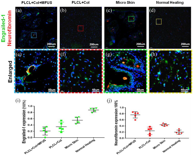
Expression of fibroblast markers, engrailied-1 (EN-1) and neurofibromin (Nf1) on rat dorsal skin tissue sections with four different treatment at day-60 post-surgery. (a–h) Immunostaining on engrailied-1 (green fluorescence) and neurofibromin (red fluorescence). (i) Semi-quantification of the engrailied-1 staining results. (j) Semi-quantification of the neurofibromin staining results. (a, e, i and j) The wound treated with tissue engineering skin (PLCL + Col + MFUS) showed that less than 20% of the cells were positively stained with engrailied-1 and more than 76% of the cells were positively stained with neurofibromin in the wound areas. (b, f, i and j) The wound treated with PLCL + Col showed about 38% of the cells were positively stained with engrailied-1, and about 28% of the cells were positively stained with neurofibromin. (c, g, i and j) The wound treated with micro skin islands showed about 50% of the cells were positively stained with engrailied-1 and about 48% of the cells were positively stained with neurofibromin. (d and h–j) The wound without treatment (Normal healing) showed more than 80% of the cells were positively stained with engrailied-1 and less than 20% of the cells were positively stained with neurofibromin.

