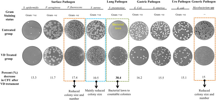
Figure 4.
VD fume affected the growth of a variety of pathogenic bacteria on agar plate. A self-explanatory picture reporting the fold changes observed in the number of colony forming units (CFUs) upon exposure of agar spread plates of different pathogenic bacteria in comparison to untreated control plates. Representative pictures of different bacterial plates in untreated and VD treated groups are provided. Type of Gram staining taken by each of the studied pathogenic bacteria is also mentioned. Mycobacterium due to waxy cell wall is incompatible with Gram staining and hence, not identified with respect to Gram staining. Categories of these pathogens based on their area of prevalence or target organ within the body, such as, surface, lung, gastric, urogenital and generic, are mentioned. Bacterial pathogens exhibiting decrease in colony size with or without decrease in CFUs are demarcated with orange and blue dashed boxes, respectively, and identified underneath with observed specific effects. Representative plate pictures of S. pneumoniae exhibiting maximum fold change in CFU is demarcated with green dashed box.

