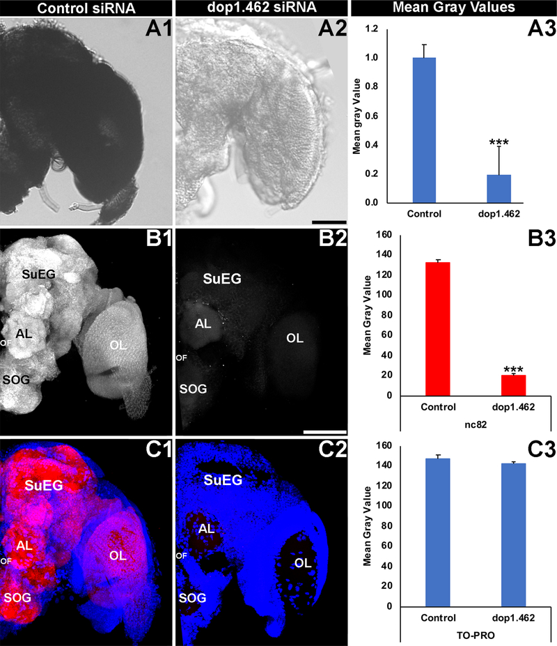
Fig. 2. Neural defects are observed in dop1.462-treated A. aegypti adults.
Broad expression of dop1.462 transcripts detected at high levels throughout the control-injected and wild-type A. aegypti adult female brain (a brain from a control siRNA-microinjected animals is shown in A1) was significantly reduced in the brains of adults injected with dop1.462 siRNA (A2; mean gray value results from three biological replicate experiments are shown in A3; n = 50 control-treated brains, and n = 60 dop1.462-treated brains). Although levels of TO-PRO nuclear staining (blue in C1, C2) were not significantly different (C3) in the brains of adults injected with control (Cl) or dop1.462 (C2) siRNA, levels of Bruchpilot (white in B1, B2; red in C1, C2), a marker of synaptic active zones (labeled by mAb nc82) were significantly reduced (B3) in the synaptic neuropil following microinjection of dop1.462 siRNA (B2, C2; compare to control siRNA treatment in B1, C1). Data were compiled from three biological replicate experiments performed on a total of 43 control-treated brains and 37 dop1.462-treated brains (B3, C3) and are represented as average mean gray values in A3, B3, and C3, in which error bars represent SEM. In all panels, brains were dissected and fixed 24 hours following injection of the mosquitoes with a 2.25 μg dose of siRNA. Student’s t-tests were used for statistical analyses of control vs. dop1.462 siRNA injected animals; ***=P<0.001 vs. control. Representative adult brains are oriented dorsal upward; scale Bar=100 μm. Labels are as follows: AL: antennal lobe; OF: oesophageous foramen; OL: optic lobe; SOG: sub-esophageal ganglion; SuEG: supra-esophageal ganglion.

