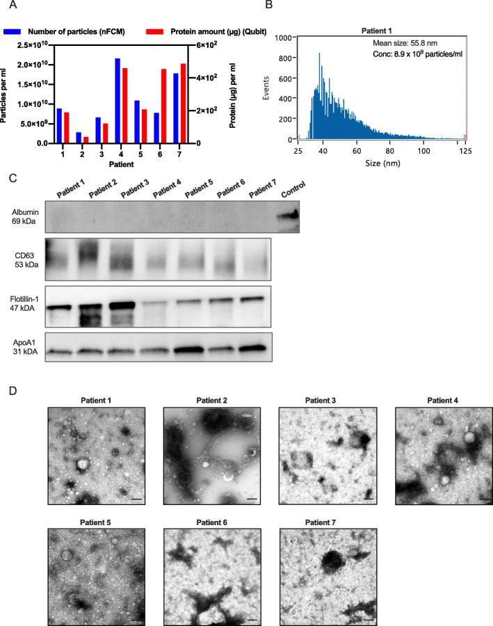
Fig. 2.
EVs isolated from lymphatic drainage fluid from breast cancer patients by SEC isolation. EVs were isolated from lymphatic drainage fluid of seven breast cancer patients using qEV SEC. EV-containing fractions were collected, pooled, concentrated using ultrafiltration and analyzed further. A EV particle number and protein amount isolated per ml of lymphatic drainage fluid from each of the 7 patients as analyzed by nFCM and Qubit. B Size distribution of EVs from patient 1 as analyzed by nFCM. C Western shows the presence of EV proteins CD63 and Flotillin and absence of albumin in EVs isolated from all patients. All EV samples contained lipoprotein ApoA1. The positive control used for albumin was human melanoma metastatic tissue and 9–10 μg protein was loaded in each lane. Western blot membranes are cropped, uncropped membranes are shown in Additional file 7. D TEM images show vesicle-like structure in the size range of 30–200 nm in EVs isolated from the breast cancer patients. Size bar, 200 nm

