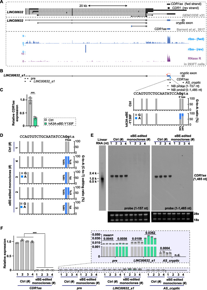
Fig. 3.
Achievement of CDR1as/ciRS-7 knockout by base editing at its 5′ back-splice site. A Schematic of the CDR1as/ciRS-7 gene organization and mapped RNA-seq signals in 293FT cells. Top, multiple transcripts were predicted/reported in the CDR1as/ciRS-7 gene locus, including a long noncoding RNA containing CDR1as/ciRS-7-residing exon (blue bar) and a cryptic exon (blank bar). Bottom, the circular molecule, CDR1as/ciRS-7, was confirmed as the major transcript produced from its gene locus, enriched after RNase R treatment. B Design of base changes at 5′ (b)ss of CDR1as/ciRS-7 by hA3A-eBE-Y130F. Top, schematic of partial CDR1as/ciRS-7 gene organization. The back-spliced CDR1as/ciRS-7 (blue bar) and a cryptic exon (blank bar) were reported to be also spliced in a long noncoding RNA. Middle, primers for RT-qPCR and probes for northern blotting. Bottom, context sequences of targeted 5′ (b)ss were shown by a, t, c, and g for intron or by A, T, C, and G for exon; G-to-A base change ratio at targeted 5′ (b)ss of back-spliced CDR1as/ciRS-7 exon was examined in transfected 293FT cell mixture. C Repression of CDR1as/ciRS-7 back-splice by base changes at its 5′ bss. RT-qPCR was performed with primers labeled in B. D Selection of monoclones with corresponding base editing changes at the 5′ bss of CDR1as/ciRS-7. Four monoclones were identified with almost 100% G-to-A base change at the exon boundary of the CDR1as/ciRS-7 5′ (b)ss, and among them, monoclones #2 and #4 have an additional G-to-A change (~ 67%) at the intron boundary of the CDR1as/ciRS-7 5′ bss. Four monoclones with unchanged bases at the 5′ bss of CDR1as/ciRS-7 were used as controls (#1 is showed in this panel). E Expression of CDR1as/ciRS-7 was undetected in the four selected monoclones with base editing changes at the 5′ bss of CDR1as/ciRS-7, revealed by northern blotting with two probes (1–157 nt and 1–1485 nt). Total RNAs were denatured and then resolved on 1.5% native agarose gel. F Back-splice of CDR1as/ciRS-7 was barely detected in the four selected monoclones with base editing changes at the 5′ bss of CDR1as/ciRS-7, revealed by RT-qPCR. Canonical splice along its cognate linear RNA was further compared by parallel RT-qPCR. n.d. indicates non-detected. C, F Error bar represents SD from three independent replicates. ∗∗∗, P < 0.001, Student’s t test

