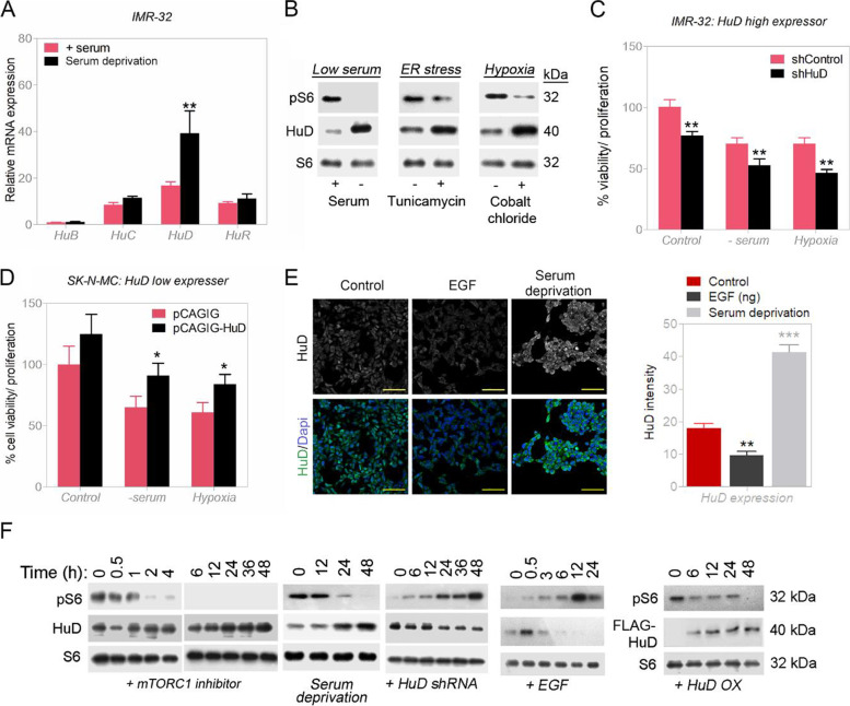
Fig. 2.
HuD levels are upregulated by stress. A Increase in HuD RNA levels by serum deprivation in IMR-32 cells. B Changes in protein HuD and pS6 levels under various forms of stress (serum starvation, ER stress and hypoxia) in IMR-32 cells by Western blot analysis. Full-length blots are presented in Supplementary Fig. S9. C Viability changes under stress condition in control or silenced HuD IMR-32 cells. D Viability changes for stressed cells (control or vector-mediated addition of HuD) in HuD low expresser SK-N-MC cells. E HuD protein changes in EGF supplemented and serum deprived cells condition by immunocytochemistry and their relative quantification. F Changes in HuD and pS6 protein following rapamycin (mTOR inhibitor), serum deprivation, silencing of HuD (HuD shRNA), vector-mediated addition of FLAG-HuD and EGF treatment by Western blot analysis. Full-length blots are presented in Supplementary Fig. S9. Data are presented as mean ± SEM; t-test: *p < 0.05, **p < 0.01, ***p < 0.001

