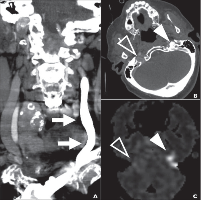
Fig. 3—
91-year-old woman with history of atrial fibrillation who was found unresponsive. NIH Stroke Scale score on arrival at emergency department was 23, and CT angiography showed basilar thrombosis (case 2).
A, CTA image obtained with left antecubital injection site to evaluate acute stroke shows florid retrograde flow of IV contrast material in internal jugular vein (arrows).
B, Axial arterial phase CTA image at skull base shows reflux of contrast material into jugular foramen (solid arrowhead) without reflux on right (open arrowhead).
C, Arterial spin-labeling MRA image obtained 24 hours after B shows pattern of hyperintensity (arrowheads) matching that in B.

