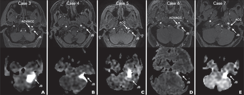
Fig. 4—
Time-of-flight (top row) and arterial spin-labeling (bottom row) MRA images show positions of jugular bulb (JB), anterior condylar vein (ACV), and anterior condylar confluence (ACC).
A, 64-year-old woman with WHO grade IV astrocytoma of left frontal lobe treated with surgery and adjuvant chemotherapy 5 years earlier presenting for routine tumor imaging (case 3).
B, 69-year-old woman with 1-year history of right-sided whooshing pulsatile tinnitus without headache (case 4).
C, 63-year-old man referred to pulsatile tinnitus clinic with episodic vertigo, nausea, headaches, and low-frequency whooshing pulsatile tinnitus (case 5).
D, 48-year-old man with vomiting, left arm clumsiness, and global weakness for 1 week and history of aortic valve replacement (case 6).
E, 74-year-old woman with new nocturnal pulsatile tinnitus (case 7).

