Fig. 5—
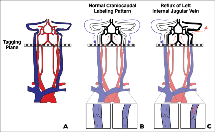
Schematic shows proposed mechanism for false localization of signal intensity to left jugular foramen on arterial spin-labeling (ASL) images.
A, In typical ASL protocols, spin-labeling is performed in cross-sectional slab (tagging plane) above carotid bifurcation.
B, With conventional flow, tagged arterial blood travels cranially to brain (red arrows) and tagged venous blood flows caudally (blue arrows), remaining below imaging plane. Inset shows valve configuration.
C, In certain patients, spin-labeled jugular blood may flow in retrograde manner to level of jugular foramen (solid red arrows) or dural sinuses (dotted red arrow), resulting in misleading appearance of arterial signal intensity. Our findings suggest that physiologic asymmetry in internal jugular vein (IJV; more frequently closed on left) contributes to reversal of flow either through increased resistance (closed valve) or from retrograde flow across incompetent left IJV valve (cases 1 and 2). Blue arrow indicates normal caudal venous blood flow. Inset shows valve configuration.

