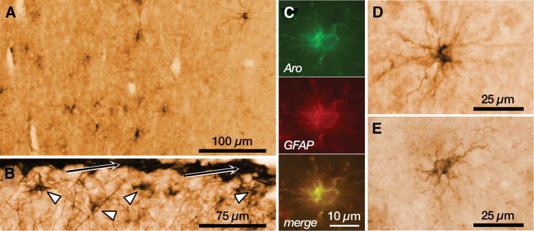
FIG. 3.
Aromatase immunoreactivity in astrocytes. (A) Panoramic view of the cortical white matter in the temporal lobe, showing immunoreactivity in cells with the morphology of fibrous astrocytes. (B) Detail of the layer 1 of the temporal cortex showing numerous immunoreactive astrocytes (arrowheads) in the proximity of the pial surface (arrows). (C) Immunofluorescence labeling of aromatase (green), the astrocyte cell marker GFAP (red), and the colocalization signal (yellow) in a cortical astrocyte. (D) Representative example of a fibrous astrocyte immunoreactive for aromatase in the cortical white matter. (E) Representative example of a protoplasmic astrocyte immunoreactive for aromatase in the cortical gray matter. All panels are from a 23-year-old male. GFAP, glial fibrillary acidic protein.

