Figure 1.
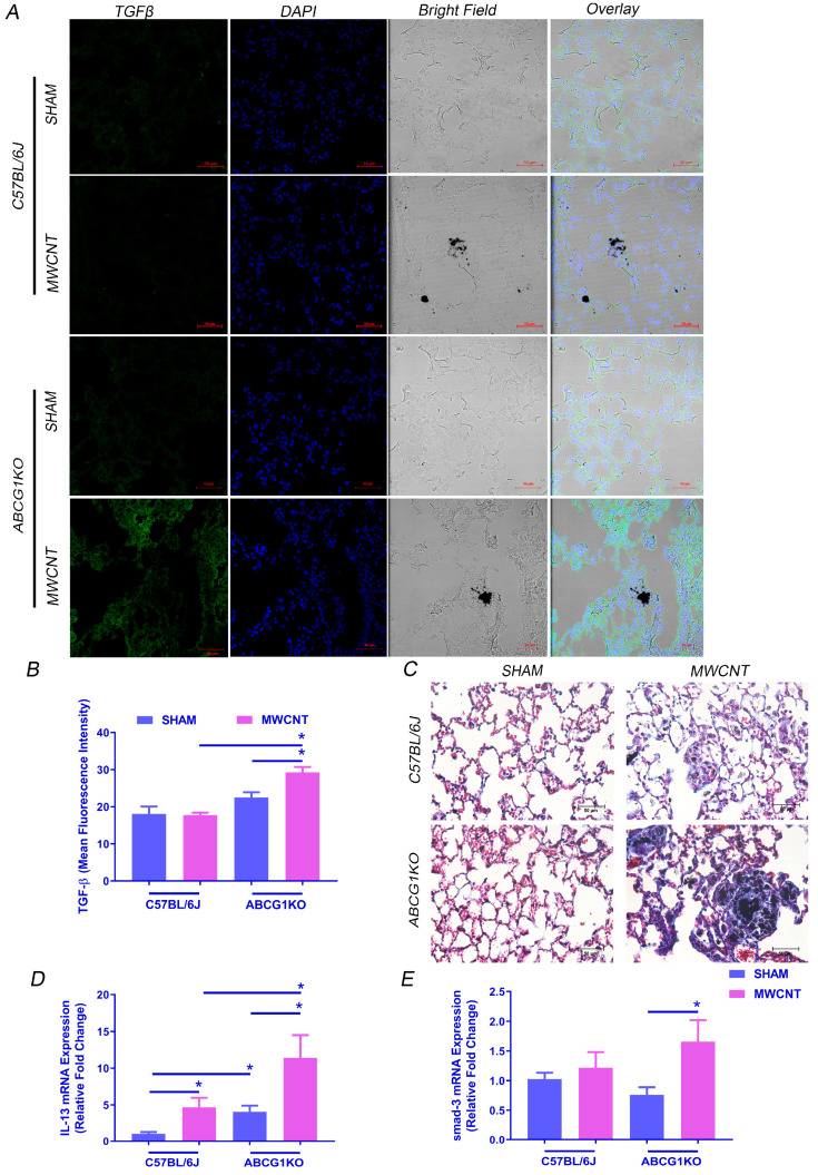
Myeloid ABCG1 deficiency increases fibrosis development and TGF-β expression in the lung of MWCNT instilled mice. (A) Representative images showing an increase in TGF-β expression in lung sections of MWCNT instilled ABCG1 KO mice. Lung sections from wild-type C57BL/6J mice or ABCG1 KO mice either sham- or MWCNT instilled were stained with TGF-β (green) and DAPI (blue). Bright field showing the presence of MWCNT in lung tissues. Scale bars: 50 μm. (B) Graphical representation of TGF-β mean fluorescence intensities using Zen 3.1 blue edition. (C) Representative light micrographs showing an increase in fibrosis in lung sections of MWCNT instilled ABCG1 KO mice using trichrome staining. Scale bars: 50 μm. (D,E) Measurement of IL-13 and smad-3 mRNA expression, respectively, in BAL cells using qRT-PCR (* p ≤ 0.05, N ≥ 3).

