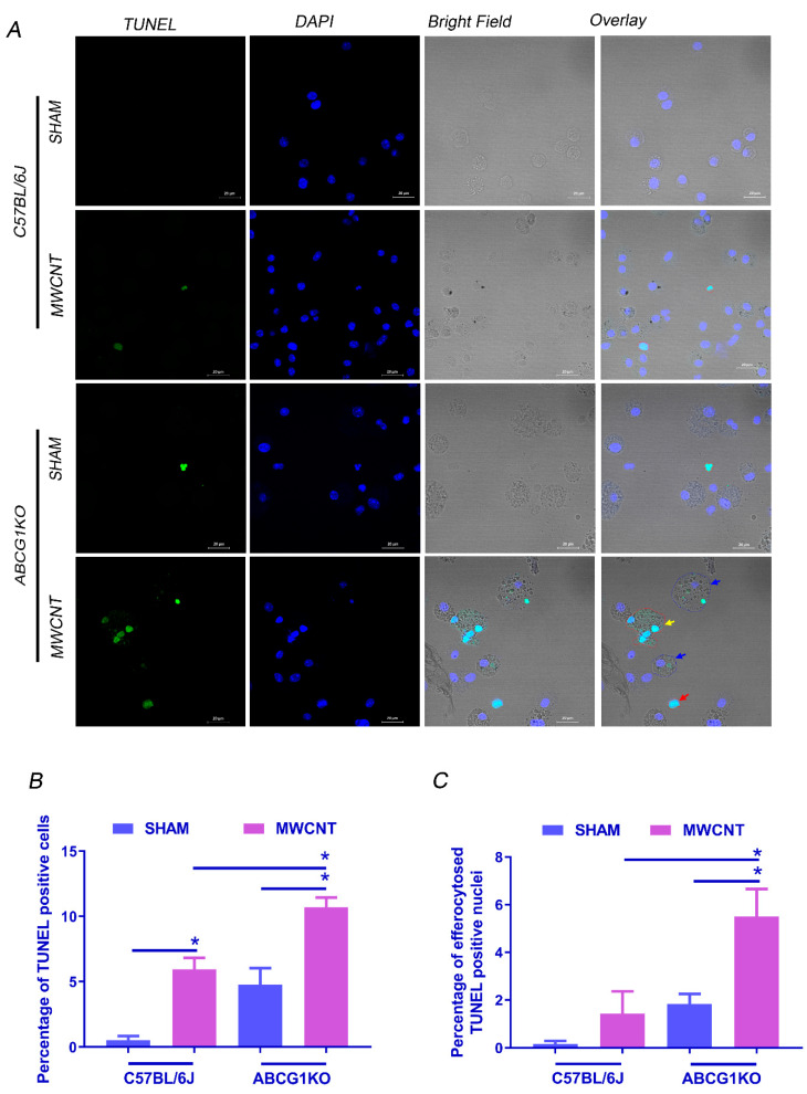
Figure 4.
Myeloid ABCG1 deficiency increases the percentage of efferocytosed apoptotic cells after MWCNT instillation. (A) Representative images showing TUNEL staining for BAL cells (40×). Cytospins of BAL cells from C57BL/6J and ABCG1 KO mice either sham or MWCNT instilled were stained with TUNEL reagent for DNA fragmentation (green) and nuclear stain, DAPI (blue). Bright field showing non-engulfed apoptotic cells (red arrow), efferocytosed TUNEL+ nuclei (blue arrow) and multi-nucleated apoptotic cell (yellow arrow). (B) Graphical representation of the percentage of TUNEL positive cells in BAL cytospins. (C) Graphical representation of the percentage of efferocytosed TUNEL positive cells. Quantification of TUNEL staining was performed blindly on 18 random 20× fields per one cytospin for each mouse. *, significant difference, p ≤ 0.05, N ≥ 3.

