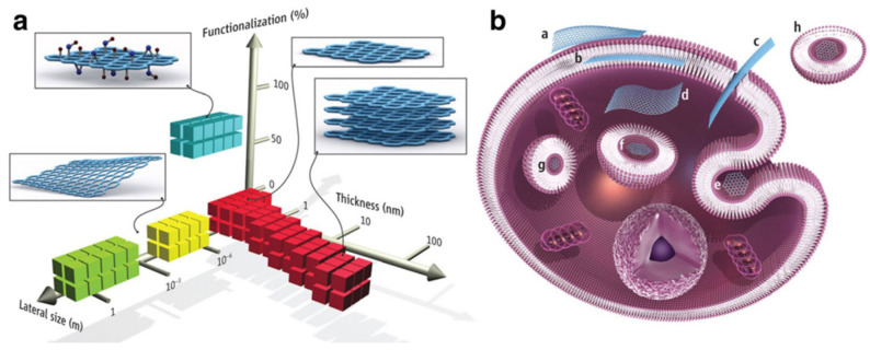
Figure 17.
(a) CBM can be described by the dimensions and surface functionalization of the material (percentage of the carbon atoms in sp3 hybridization). Green squares represent epitaxially grown graphene; yellow, mechanically exfoliated graphene; red, chemically exfoliated graphene; blue, GO. (b) Possible interactions between CBM with cells (a) Adhesion onto the outer surface of the cell membrane. (b) Incorporation in between the monolayers of the plasma membrane lipid bilayer. (c) Translocation of the membrane. (d) Cytoplasmic internalization. (e) Clathrin-mediated endocytosis. (f) Endosomal or phagosomal internalization. (g) Lysosomal or other perinuclear compartment localization. (h) Exosomal localization. The biological outcomes from such interactions can be considered to be either adverse or beneficial, depending on the context of the particular biomedical application. Different CBM will have different preferential mechanisms of interaction with cells and tissues that largely await discovery. Reproduced from Ref. [135]. Copyright 2014 the American Association for the Advancement of Science.

